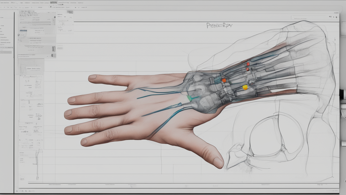
In the world of medical diagnostics, advancements in technology have always played a crucial role in improving patient outcomes. One area that has seen remarkable progress is the field of fracture detection, where artificial intelligence (AI) is revolutionizing the way X-rays are interpreted and diagnoses are made.
Recent studies have highlighted the potential of AI in enhancing patient treatment by enabling early and accurate diagnoses, which are crucial for preventing adverse outcomes. Deep learning algorithms have been developed to identify various conditions in wrist X-rays, including wrist fractures, benign bone tumors, and bone necrosis. These AI technologies offer valuable support in emergency departments and for physicians who may not specialize in interpreting hand or wrist X-rays.
Dr. Jorma Ryhänen, the head of hand surgery at HUS, and his research team have developed an AI model that can accurately identify 97% of distal radius fractures, which constitute 20% of all emergency department fractures. This is a significant achievement, as misinterpretation of X-rays is common and can lead to unnecessary delays or complications in treatment. Dr. Ryhänen explains, “The AI model we’ve developed excellently identifies these fractures, potentially offering immediate recommendations for treatment, whether cast immobilization or surgery.”
The benefits of AI in emergency departments extend beyond fracture detection. Enchondroma, a common benign bone tumor in the hand, can often go unnoticed in emergency situations, especially when fractures are present. Dr. Ryhänen’s team has developed an AI model that can identify enchondromas in the hand, proving to be a useful tool for emergency department physicians who may be less familiar with hand-related ailments. Dr. Turkka Anttila, a hand surgeon and member of the research team, shares, “The detection of enchondromas is an exciting first step towards automated diagnostics of bone tumors in the hand area.”
In another study, the AI model developed by Dr. Ryhänen’s team demonstrated its ability to identify lunate necrosis in X-rays, a rare condition that mainly affects men aged 20–40. Early stages of necrosis are often invisible to the human eye, leading to delayed diagnoses and limited treatment options. However, the AI model was able to identify necrosis in 28 out of 30 cases, surpassing a group of experienced specialists in accuracy.
These advancements in AI technology are setting the stage for a future where medical imaging and diagnostics are transformed. Dr. Ryhänen believes that AI algorithms will significantly change patient diagnostics and treatment in surgical practice soon. He explains, “AI can tirelessly and cost-effectively sift through large volumes of images, identifying abnormalities, risk factors, and complications, thus speeding up and improving treatment outcomes.”
However, the development and validation of reliable and generalizable AI models require high-level interdisciplinary expertise and research. Dr. Ryhänen’s research team has several promising ideas for further investigation, aiming to push the boundaries of AI’s capabilities in medical diagnostics.
As AI continues to evolve and make strides in the field of fracture detection, the future looks promising for improving patient outcomes. The ability to accurately and swiftly interpret X-rays can significantly reduce misdiagnoses, unnecessary delays, and complications in treatment. The integration of AI technologies in emergency departments and medical practices holds the potential to transform the way fractures are diagnosed and treated, saving precious time and lives in the process.


Use the share button below if you liked it.